|
|
|


/
 0 0/
/
/


|


| 상품번호 : 232864 | |||||||
|
|||||||


 |
 (0) (0) |
 (0) (0) |
 |
|---|
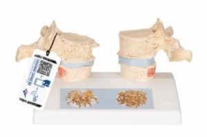
● 골다공증과 정상 흉추를 비교하기 위한 인상적인 교육용 모형 입니다. ● 의학 연구 및 환자 상담에 이상적입니다. ● 11번째와 12번째 흉추를 보여줍니다. ● 더 좁은 추간판을 가진 순차 골다공증 흉추의 복제품은 스탠드의 왼쪽에 있습니다. ● 위쪽 척추뼈가 가운데로 나뉘어져 있습니다. ● 자석으로 부착된 척추 반쪽은 절단면을 표시하기 위해 쉽게 분리할 수 있습니다. ● 이것은 소결로 인한 골절된 척추체 상부의 명확한 시각화를 가능하게 합니다. ● 골극으로 나타나는 뼈의 퇴행성 변화도 확인할 수 있습니다. ● 비교를 위해 오른쪽에 추간판이 있는 두 개의 해당하는 건강한 척추의 복제물이 제공됩니다. ● 상부 척추체의 절반은 자력으로 부착되어 있으며 분리할 수 있습니다. ● 베이스의 디테일 일러스트는 뼈 생검에서 얻은 2개의 3D 마이크로 CT 영상을 나타냅니다. ● 건강한 뼈에 비해 골밀도가 낮은 골다공증성 뼈의 미세구조를 나타낸 것입니다.
● Impressive didactic model for comparing osteoporotic and normal thoracic vertebrae.
● Ideal for medical studies and patient consultation.
● The 11th and 12th thoracic vertebrae are shown.
● Reproductions of sequential osteoporotic thoracic vertebraewith narrower intervertebral disc are located on the left of the stand.
● The upper vertebra is divided in the middle.
● The magnetically attached vertebral half can be removed easily to show the cut surfaces.
● This allows clear visualization of the fractured upper part of the vertebral body caused by sintering,
i.e. collapse of the bony substance in the course and as a result of osteoporosis.
● Degenerative changes in the bone, manifested as osteophytes, are also identifiable.
● For comparison, reproductions of two corresponding healthy vertebrae with intervertebral disc are provided on the right side.● One half of the upper vertebral body is magnetically attached and can be removed.
● A detail illustration on the base depicts two 3D micro CT images obtained from bone biopsies.● These illustrate the microacrchitecture of the osteoporotic bone, which has a lower bone density compared to healthy bone.
 |
(0) |
(0) |
|
|---|
 |
|
|
(0) |
(0) |
|
|---|
 |
|
2. 해외구매 특성상 주문에서 배송까지는 평균 10~15일이 소요됩니다. 간혹 현지 제품 수급에 따라 부득이하게 시일이 더 소요 될 수 있으니 구매시 좀 더 여유있게 주문하시길 권합니다.
3. 해외 내수품인 관계로 A/S에 대해서는 별도의 책임을 지지 않습니다.
4. 해외배송 특성상 주문접수후 배송상태가 배송준비중으로 넘어간 경우 해외에서 국내로의 배송이 이루어지고 있다는 뜻입니다. 따라서 배송준비중으로 배송상태가 넘어간 경우 취소및 반품이 불가하므로 이점 양해 부탁드립니다.
5. 타 해외구매대행 사이트에서 주문하신 물건과 주문날짜가 겹치지않도록 주의해 주십시오. 통관날짜가 같을 경우 합산관세가 부가되게 됩니다.
|


|