|
|
|


/
 0 0/
/
/


|


| 상품번호 : 232959 | |||||||
|
|||||||


 |
 (0) (0) |
 (0) (0) |
 |
|---|
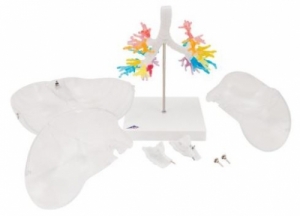
● 이 독특한 후두 CT 기관지 분지 모형은 인간(남성, 약 40세)의 컴퓨터 단층 촬영 데이터를 기반으로 생성되었습니다. ● 이 절차의 특별한 점은 자연스러운 공간 3D 관계와 분절 기관지의 상호 위치를 보존하고 사실적으로 입증할 수 있다는 것입니다. ● 결과는 실물과 같은 CT 기관지 및 후두입니다. ● 이 모형은 인간 폐의 해부학적 구조를 연구하는 독특한 방법입니다. ● 설골과 후두개가 있는 후두와 일차 및 소엽 기관지가 있는 기관은 기관지 분지 모형에서 한 가지 색상으로 묘사됩니다. ● 후두는 두 번째 기관 연골 수준에서 분리 가능하고 정중면에서 분할 가능합니다. ● 기관지와 후두의 후두개가 유연하게 장착됩니다. ● 다양한 분절 기관지는 신축성 있는 재질로 되어 있으며 시각적으로 구별하기 쉽도록 다양한 투명 색상으로 표현되어 있습니다. ● 투명한 폐는 기관지와 후두에서 분리 가능합니다.
● This unique CT bronchial tree model with larynx was created on the basis of computer tomography data of a human
(male, approx. 40 years).
● What is special about this procedure is that the natural spatial 3D-relations and the reciprocal location
of the segmental bronchi can be preserved and demonstrated in a realistic way.
● The result is a life-like CT bronchial tree and larynx.
● This model is a unique way to study the anatomy of the human lungs.
● The larynx with hyoid bone and epiglottis and the trachea with primaryand lobar bronchi are depicted in one color on the bronchial tree model.
● The larynx is detachable at the level of the second tracheal cartilage and divisible in the median plane.
● The epiglottis on the bronichal tree and larynx is mounted flexibly.
● The various segmental bronchi are made of elastic material and depictedin various transparent colors so that they are easier to distinguish visually.
● The transparent lungs are detachable from the bronchial tree and larynx.
 |
(0) |
(0) |
|
|---|
 |
|
|
(0) |
(0) |
|
|---|
 |
|
2. 해외구매 특성상 주문에서 배송까지는 평균 10~15일이 소요됩니다. 간혹 현지 제품 수급에 따라 부득이하게 시일이 더 소요 될 수 있으니 구매시 좀 더 여유있게 주문하시길 권합니다.
3. 해외 내수품인 관계로 A/S에 대해서는 별도의 책임을 지지 않습니다.
4. 해외배송 특성상 주문접수후 배송상태가 배송준비중으로 넘어간 경우 해외에서 국내로의 배송이 이루어지고 있다는 뜻입니다. 따라서 배송준비중으로 배송상태가 넘어간 경우 취소및 반품이 불가하므로 이점 양해 부탁드립니다.
5. 타 해외구매대행 사이트에서 주문하신 물건과 주문날짜가 겹치지않도록 주의해 주십시오. 통관날짜가 같을 경우 합산관세가 부가되게 됩니다.
|


|